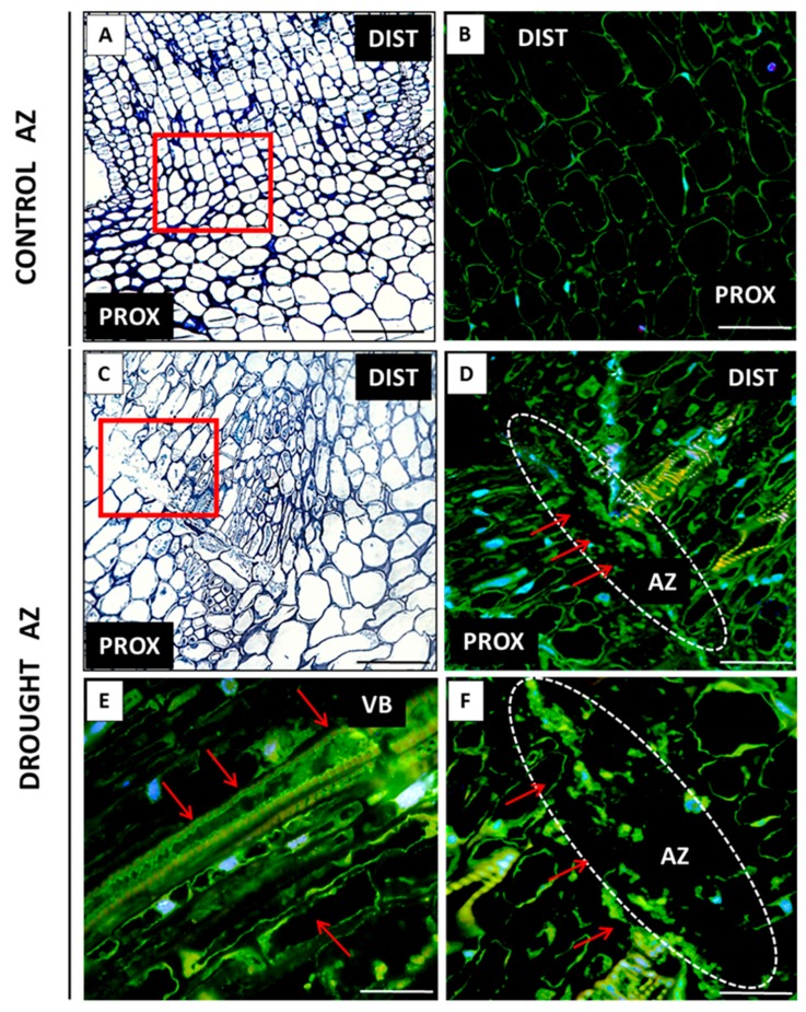
Figure 7.
Tissue and subcellular localization of abscisic acid (ABA) in the abscission zone (AZ) of Lupinus luteus flowers grown in drought conditions (D–F) and in the AZ of control plants (B). The AZs were excised on the 48th day of development. Control plants were cultivated under optimal soil conditions (70% WHC). Part of plants was subjected to drought conditions for 2 weeks (25% WHC). The AZ regions are indicated by white curves (D,F). The presence of ABA (D–F) is highlighted by red arrows. Image F is magnified D region. Image E corresponds to the vascular bundles’ area. DAPI was used for nuclei staining. The examined regions of AZs used for the immunofluorescence studies are indicated by red squares (A,C). Abbreviations: PROX—stem fragments below the AZ, DIST—flower pedicel fragments above the AZ, VB—vascular bundles. Scale bars: 100 µm (A,C,D), 60 µm (B), 40 µm (E,F).

