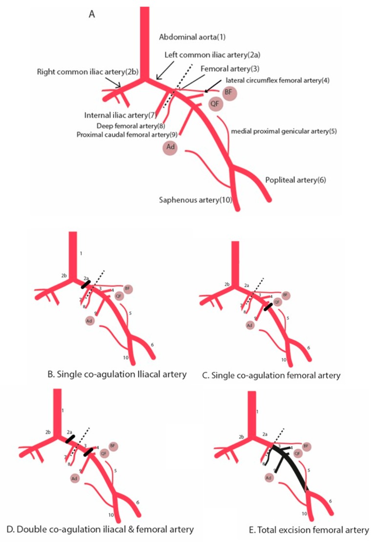
Figure 1.
Schematic diagram illustrating the hind limb ischemia and different surgical methods to induce hind limb ischemia. (A) Hind limb vasculature. (B) Single electrocoagulation of the iliac artery. (C) Single electrocoagulation of the femoral artery. (D) Double electrocoagulation of the iliac and femoral artery. Alternatively, the electrocoagulation of the iliac artery can be replaced by a second electrocoagulation of the femoral artery just above the bifurcation of the saphenous and popliteal artery. (E) Total excision of the femoral artery. Round circles are muscle groups: (Ad) adductor muscle group. (QF) Quadriceps femoris. (BF) Biceps femoris. Dotted line is the inguinal ligament. Black beam is the occlusion site.

