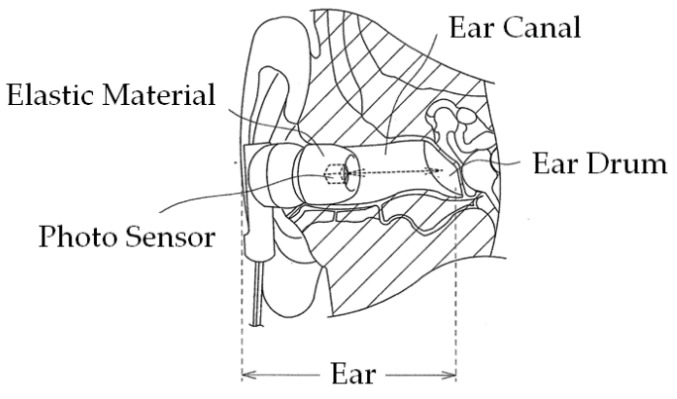
Figure 2.
Principle of ear canal movement measurement using ear sensor. Occlusion is performed by the temporalis and masticatory muscles, including the masseter muscle and the temporomandibular joint. Occlusion causes a change in the ear canal shape near the masticatory muscles and the temporomandibular joint. The ear sensor measures this shape change in the ear canal during occlusion optically and noninvasively. A small photosensor is attached to the ear sensor. This photosensor houses a light-emitting diode (LED) with an emission wavelength of 940 nm and a phototransistor, as illustrated in Figure 1. The ear sensor irradiates the skin of the ear canal with infrared light, and the reflected light is then received by the phototransistor to measure the change in the ear canal shape.

