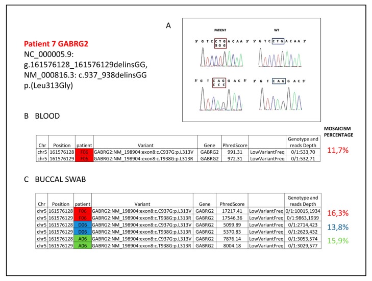
Figure 3.
(A) Patient electropherograms show the mosaic double nucleotide substitution in the first and second position of the CTG codon (framed) with the mutated GGG codon (framed) replacing the aminoacid Leu at position 313 of the GABRG2 gene with Gly. (B) Annovar table revealing the low-rate (12%) mosaicism for the double base substitution in DNA from peripheral blood. (C) Nextera-XT-Library-prep protocol performed with three different pairs of primers (red-blue-green) shows on DNA from buccal swab a mosaic mutation percentage comparable to that obtained on blood.

