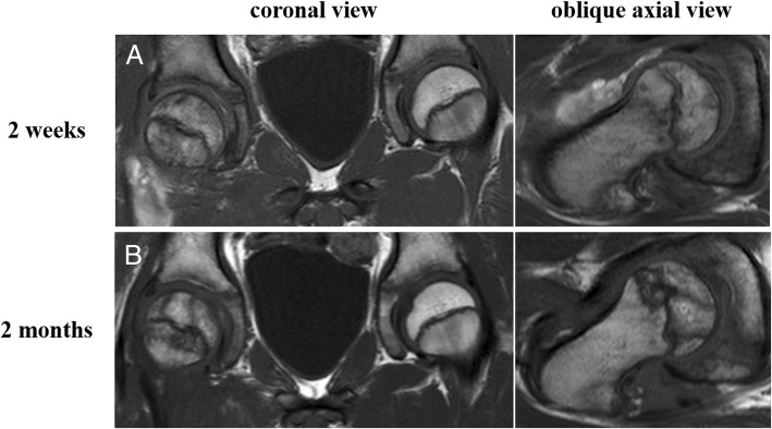
Fig. 4.
Coronal and oblique axial T1-weighted images at 2 weeks (a) after injury show diffuse area with low signal intensity in the proximal femur suggesting ischemia. Coronal and oblique axial T1-weighted images at 2 months (b) after injury show two bands with low signal intensity in the epiphysis of the femoral head, suggesting osteonecrosis

