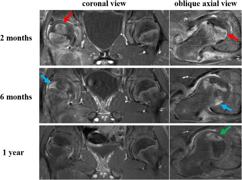
Fig. 5.

Coronal and oblique axial serial gadolinium-enhanced magnetic resonance images (MRIs) obtained at 2 months, 6 months, and 1 year. MRI at 2 months shows gadolinium enhancement in the central region (red arrows) and nonenhancement in the peripheral region of the femoral capital epiphysis. MRI at 6 months shows gadolinium enhancement spreading from the center toward the lateral and posterior regions of the femoral head (blue arrows). MRI at 1 year shows femoral head intensity equivalent to that on the contralateral side except for anterior region with slight collapse of articular surface of the femoral head (green arrow)
