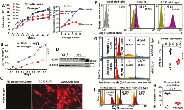
Figure 3.
Proliferation and metabolic activity of adipose-derived stem cells (ADSC) in KL-/- and wild-type animals. (A) Proliferation and growth curve of ADSC: Cells were counted daily in P3, and their cumulative cells number was recorded until senescence in P12, in KL-/- and wild-type group. (B) 5 × 10³ cells/well were seeded and metabolic activity was measured by MTT assay. (C) Representative images showing morphology of P3 ADSC in KL-/- and wild-type mice. (D) Western blot of Ki-67 antigen (a nuclear protein and cellular proliferation marker). (E) Flow cytometric analysis of Ki 67, (F) and % comparative representation of Ki-67 in KL-/- and wild-type ADSC. (G) Cell cycle analysis was performed to determine healthy versus unhealthy ADSC, (H), which showing higher proapoptotic population in % for KL-/- group compared to wild type. (I) Flow cytometric analysis was carried out to monitor the reactive oxygen species (ROS), in KL-/- versus wild-type ADSC and ROS inducer (Pyocyanin 1 µM) was used to generate positive control. mean ± SEM, statistically significant values are: *p = .05; **p = .01; ***p = .001, scale bar: 200 μm.

