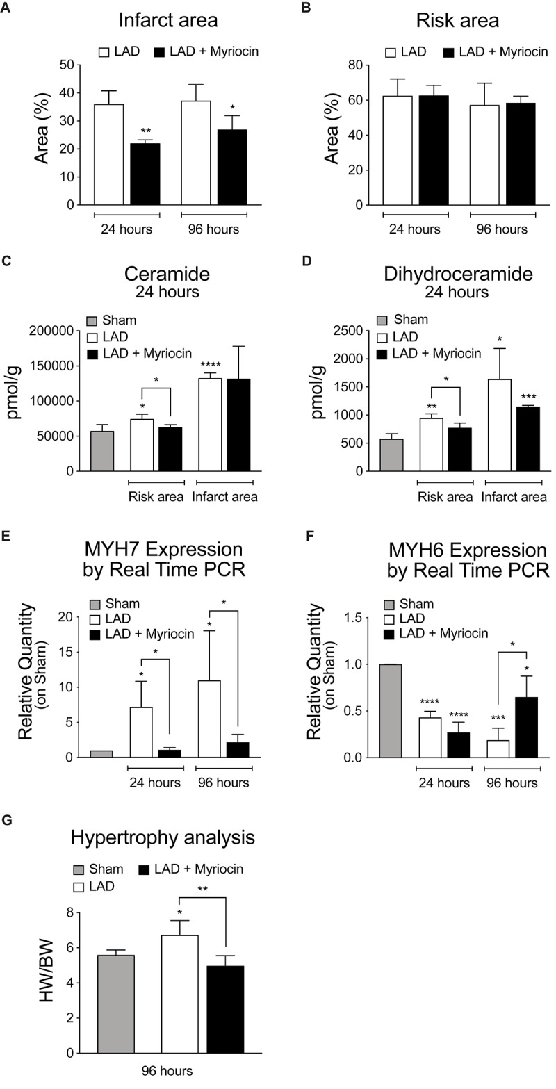
FIGURE 1.

Analysis of SLN/myr treatment effects on infarct areas, sphingolipid content and hypertrophy. (A) analysis of the infarct area (expressed as percentage on at-risk area, on the left) and (B) of the area at-risk (expressed as percentage on total tissue, on the right) in infarcted myocardium, 24 h and 96 h after surgery. (C) LCMS measurement of ceramides and (D) dihydroceramides species in infarct and at-risk area, 24 h and 96 h after surgery. qRT-PCR analysis of (E) β-myosin heavy chain (MYH7) and (F) α-myosin heavy chain (MYH6), 24 h and 96 h after surgery, expressed as relative quantity versus sham group. (G) measurement of myocardial hypertrophy, 96 h after surgery, calculated as ratio between heart weight on body weight. All data are expressed as mean ± SD. Statistical significance refers to I/R and SLN/myr treated I/R groups as compared to sham animals. The comparison between I/R and SLN/myr treated I/R groups is indicated by the connecting line (*p < 0.05; ∗∗p < 0.01; ∗∗∗p < 0.001; ****p < 0.0001). Sham animals: gray bar; I/R group: white bar; SLN/myr treated I/R group: black bar.
