
Fig. 1.
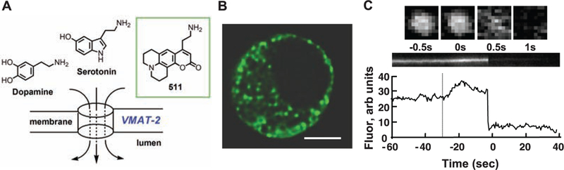
In mouse chromaffin cells, FFN511 is accumulated in LDCVs and is released by exocytosis. (A) Chemical structure of FFN511. (B) Multiphoton image of a chromaffin cell reveals a distribution of FFN511 that is consistent with LDCVs. Scale bar, 5 μm. (C) FFN511 exocytosis from a LDCV observed with TIRFM images acquired at 500 ms intervals. The upper row shows consecutive images of a single vesicle. Orthogonal section through this vesicle and its integrated intensity are in the middle and lower panels. The dotted line indicates stimulation by high potassium; the delay after stimulation is typically observed in this preparation (movie S1).