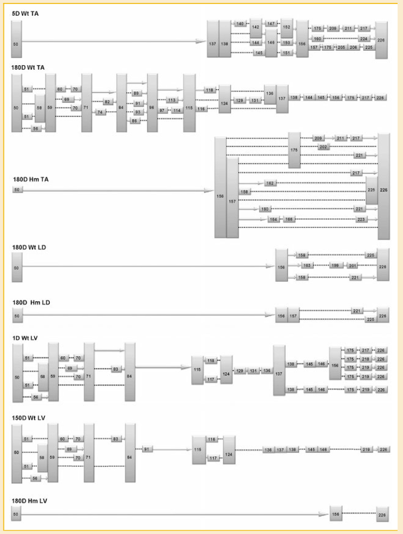
Fig. 4.

Summary of the exon profile of titin mRNA corresponding to middle Ig and PEVK region in different tissues. The colored boxes are exons (with their numbers), the solid arrows indicate consecutive exons. The dotted lines denote direct attachment between the adjacent numbered exons, the information between exon 156 to 226 in 180D Hm LV is missing.
