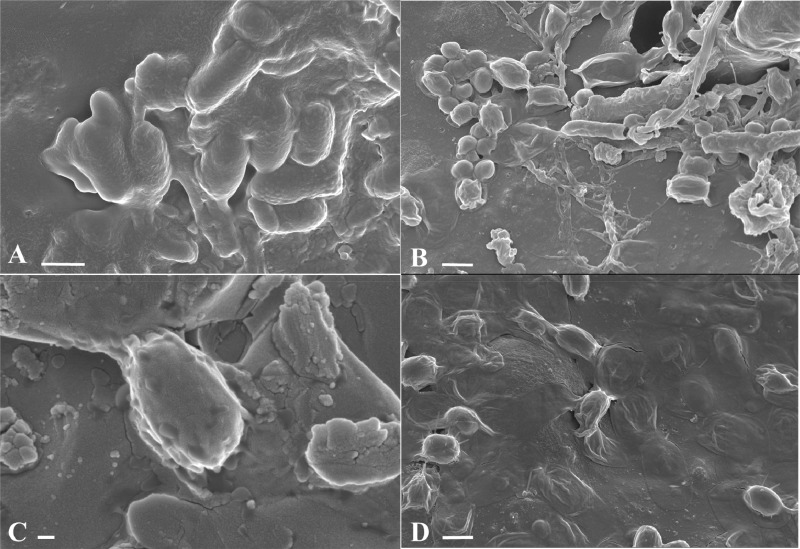
FIG 5.
Scanning electron micrographs of C. difficile spores present on spiked hospital stainless steel and floor vinyl before and after treatment with NaDCC at 1,000 ppm for 10 min. Images are NaDCC-treated DS1813 P spores on stainless steel (A), DS1748 U spores on floor vinyl (B), DS1748 U spores on stainless steel (C), and R20291 U spores on floor vinyl (D). Arrows highlight areas in the exosporium layer. Scale bars are 1 μm in panels A, B, and D and 200 nm in panel C.

