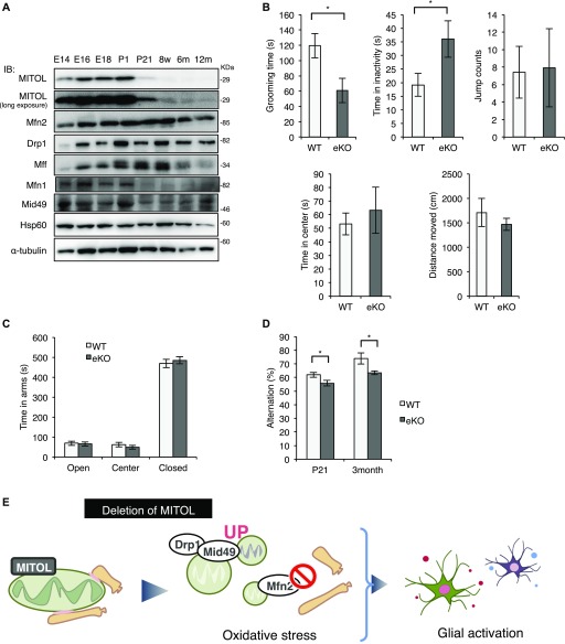
Figure 5. MITOL KO mice exhibit mild developmental disorder.
(A) High expression of MITOL in the developing neurons. Immunoblot analysis on whole cell lysates from the mouse cerebral cortex at each developmental stage with the indicated antibodies. (B) Reduced grooming time and increased immobility time in eKO at P21 in the open field test (n = 17 for WT and n = 12 for eKO; *P < 0.05, t test). (C) No obvious change between WT and eKO in the elevated plus maze test. (n = 16 for WT and n = 17 for eKO at P21; *P < 0.05, t test). (D) Reduced spontaneous alternation in eKO in the Y-maze test (n = 16 for WT and n = 17 for eKO at P21, n = 7 for WT and n = 5 for eKO at 3-mo old mice; *P < 0.05, t test). All error bars indicate SEM. (E) Schematic model for the role of MITOL in vivo. MITOL induces the formation of ER-mitochondria contact sites by Mfn2 activation and promotes CL biosynthesis partly through ER-mitochondria contact sites-dependent efficient uptake of lipids from the ER to mitochondria. In addition, MITOL blocks excessive mitochondrial fission mediated by Mid49 and Drp1. Collectively, MITOL maintains mitochondrial network including ER tethering. These functions of MITOL in developing neurons prevents oxidative stress-induced glial activation, therefore, dysfunction of MITOL confers a risk for psychiatric disorders. E, Embrionic day; m, Month-old; P, Postnatal day; w, Week-old.

