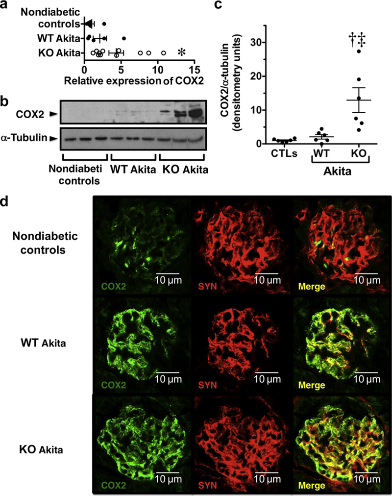
Figure 7 |. Effect of diabetes and transient receptor potential cation channel C6 (TRPC6) knockout (KO) on calcineurin target genes.

(a) Expression of cyclooxygenase 2 (COX2) mRNA tended to increase in both groups of Akita mice compared with nondiabetic controls, and this difference was statistically significant for the KO Akita group. (b,c) Expression of the COX2 protein was significantly increased in KO Akita mice compared with either nondiabetic controls or wild-type (WT) Akita mice. (d) Tissue sections were stained for COX2 (green) and the podocyte marker synaptopodin (SYN; red) and examined by confocal microscopy. In nondiabetic controls, focal areas COX2 staining were observed, which largely colocalized with the podocyte marker SYN. In both WT and KO Akita mice, COX2 staining also colocalized with the podocyte marker SYN but was more prominent and detected diffusely in the glomerular areas compared with nondiabetic controls. The intensity of staining tended to be more prominent in KO Akita mice, consistent with immunoblotting and quantitative real-time polymerase chain reaction studies. Data for real-time polymerase chain reaction, immunoblotting, and immunofluorescence studies were similar in nondiabetic KO and WT mice (nondiabetic controls), and these data were combined for statistical analyses. For immunofluorescence studies, 4–5 mice were studied per group. *P < 0.05 or †P < 0.025 versus nondiabetic controls, ‡P < 0.05 versus WT Akita mice. CTL, controls. To optimize viewing of this image, please see the online version of this article at www.kidney-international.org.
