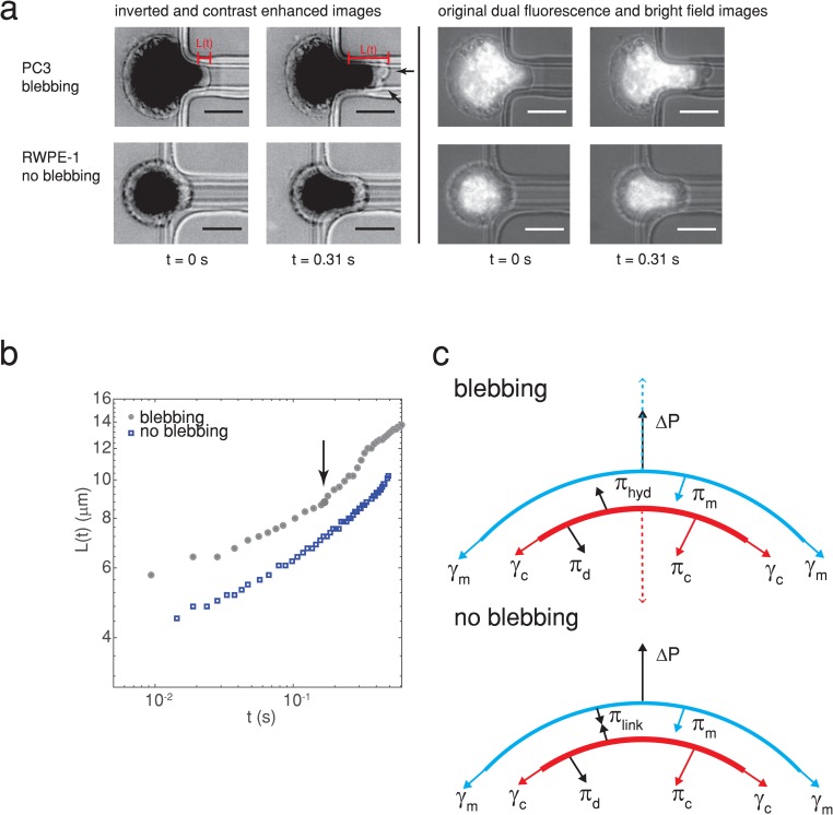
FIG. 1.
(a) A highly metastatic PC3 prostate cancer cell forms plasma membrane blebs when flowing into a constricted microfluidic channel (upper panel), while a normal RWPE-1 prostate cell does not (lower panel). Arrowheads indicate blebs. The images on the left are inverted and contrast enhanced (grayscale values in the range of [0 128] were stretched to [0 255]), and nuclei were fluorescently stained with Hoechst 33342. Scale bars represent . The original unenhanced dual fluorescence and bright-field images are shown on the right. The pressure drop across the cell is 1000 Pa. The time when the cell first enters the channel is determined from videography. The cell extension length is measured from images such as these and used to calculate rheological creep . The power law fits to vs give the cells’ stiffness and fluidity (see the main text for details). (b) The temporal change in the cell extension length for a blebbing and nonblebbing cell [both from (a)], where an arrow indicates the onset of bleb formation. (c) Simple model of the forces exerted on the plasma membrane, actin cortex, and membrane-cortex linkers for a nonblebbing (no blebbing, bottom) and a blebbing cell (top). The main forces for a nonblebbing cell are the pressure drop across the cell , the membrane tension resulting in pressure , the cortical tension resulting in pressure , the force exerted on the plasma membrane and cortex by linker proteins resulting in pressure , and the pressure resulting from viscous dissipation and friction . Arrows indicate the direction that the forces act. For a sufficiently high distribution of linkers and low pressure drop (typically 10), the plasma membrane does not become detached from the actin cortex. When is sufficiently large to detach the linker proteins, the actin cortex moves inwards toward the center of the cell due to cortical tension as denoted by the dashed red arrow, driving cytoplasmic fluid toward the plasma membrane resulting in a hydrodynamic pressure . This causes a bleb to form, pushing the plasma membrane outwards as depicted by the blue dashed arrow.10,33,34

