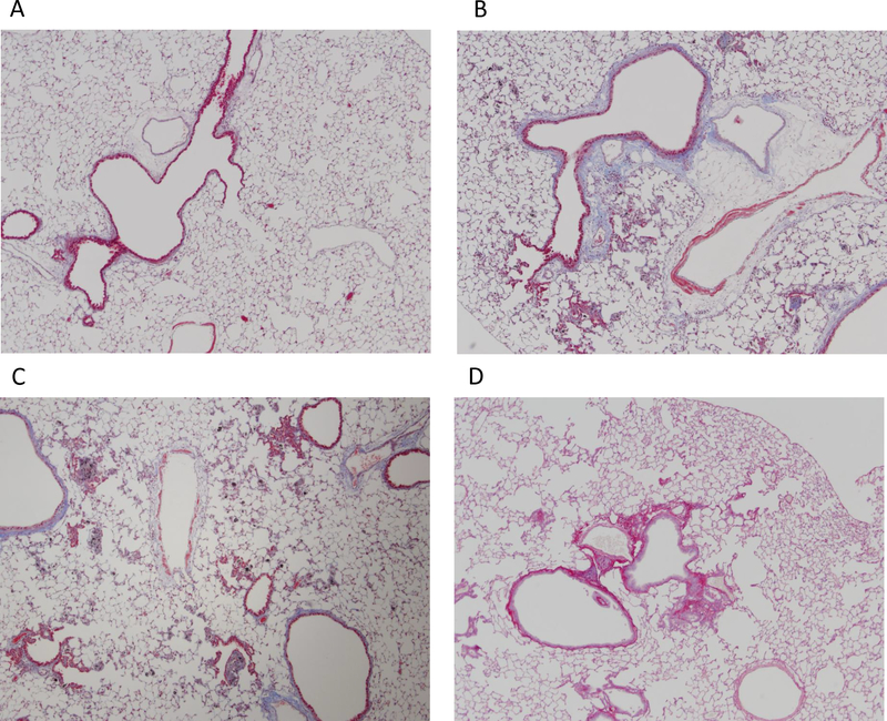
Figure 2.
Left lung trichrome and Sirius red pathology staining at 1 year. Lung sections were evaluated by visualization of collagen at 4× magnification. (A) DM (negative control) trichrome staining; (B) mild fibrosis with focal emphysema, 40 μg MWCNT, trichrome staining; (C) mild fibrosis with focal emphysema, 80 μg MWCNT, trichrome staining; (D) mild fibrosis, 120 μg asbestos, Sirius red staining.

