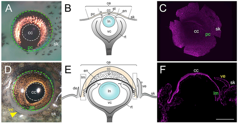
Fig. 1.

Larvae (stage 50–52) and adult (stage 66) Xenopus eyes. (A) Tadpole eye showing regions of the cornea. Region enclosed by white dotted line represents the central cornea (cc), green dotted line represents the region enclosing the peripheral cornea (pc), and the pigmented skin epithelium (sk) lies outside this line. (B) Schematic drawing of the larval Xenopus eye. The corneal epithelium is shaded light yellow, while the skin is shaded light grey. (C) Hoechst labeling in a larval corneal whole-mount. (D) Adult Xenopus eye. White dotted line encloses the central cornea (cc), green dotted line encloses the limbal region (lm), and yellow arrowhead points to the ventral eyelid (ve). The pigmented skin (sk) surrounds the eye. (E) Schematic drawing of the adult frog eye. The corneal epithelium is shaded light yellow, while the eyelid and skin is shaded light grey. (F) Hoechst labeling in a cross-section of adult Xenopus cornea, (a) apical surface of corneal epithelium, (b) basal surface of corneal epithelium, (ce) corneal epithelium, (de) dorsal eyelid, (en) corneal endothelium, (i) inner portion of eyelid, (In) lens, (o) outer surface of eyelid, (on) optic nerve, (rt) retina, (st) corneal stalk, (sr) stroma, (vc) vitreous chamber. Scale bar in F equals 470 μm for A, 520 μm for C, 800 μm for D, and 400 μm for F. (For interpretation of the references to colour in this figure legend, the reader is referred to the Web version of this article.)
