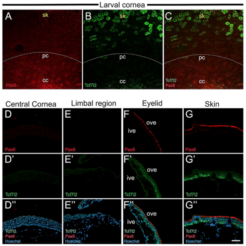
Fig. 11.

Larval and adult frog epithelia immunostained with Pax6 and Tcf7l2 antibodies. (A–C) Lower magnification confocal images of larval whole-mount cornea showing Pax6 (red), and Tcf7l2 (green) co-staining. The white dotted line represents the boundary demarcating the cornea and skin. (A) A gradient of Pax6 expression is higher in the central cornea (cc), and is lost progressively towards the peripheral cornea (pc), and skin (sk). (B) Tcf7l2-positive cells are abundant in the skin region. (C) Merged image showing Pax6 and Tcf7l2 expression. (D-G, D′-G′ and D”-G″) Cross-sections of adult frog cornea, eyelid and skin immunolabeled with Pax6 (red) and Tcf7l2 (green). The apical surface is located towards the top of each image, and the basal surface towards the bottom. D″, E″, F″ and G″ are merged images for D, D’; E, E’; F, F′ and G, G′, respectively, with Hoechst labeled nuclei (blue). (D, D′ and D″) No Pax6 or Tcf7l2 expression is detected in the central cornea. (E, E′, and E″) The limbal region has undetectable Pax6 or Tcf7l2. (F, F′ and F″) Ventral eyelid region showing Pax6 and Tcf7l2 expression. The inner surface of the eyelid does not stain for either antibody. Outer surface of the eyelid has Pax6 in the apical cells, and Tcf7l2 mainly in the basal layer of cells. (G, G′ and G″) Skin of the adult frog has Pax6 in apical cells of the epithelium, and Tcf7l2 in the basal and intermediate layers of cells, cc, central cornea; ive, inner ventral eyelid; ove, outer ventral eyelid; pc, peripheral cornea; sk, skin. Scale bar in G″ equals 60 μm for A-C, 50 μm for D-G, D′-G′, D”-G″. (For interpretation of the references to colour in this figure legend, the reader is referred to the Web version of this article.)
