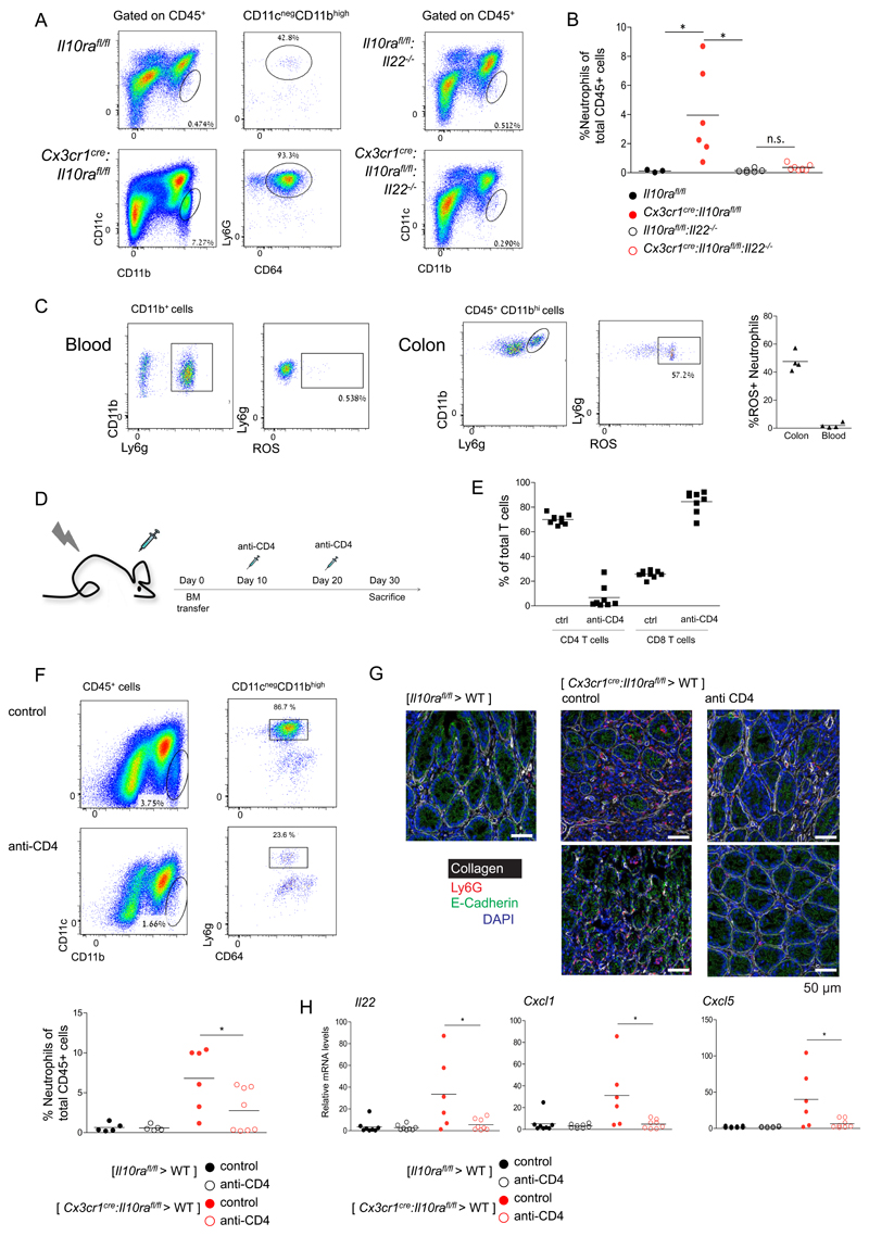
Figure 6. Neutrophil recruitment to the colonic lamina propria depends on CD4+ T cells.
(A) Representative plots of flow cytometry analysis of the colonic lamina propria of indicated mouse strains.
(B) Quantification of flow cytometry analysis according to gating strategy indicated in A.
(C) Representative plots of flow cytometry analysis of the colonic lamina propria of Cx3cr1cre:Il10rafl/fl mice (left). Quantification of flow cytometry analysis (right).
(D) Schematic of T cell depletion protocol
(E) Quantification of flow cytometry analysis of mesenteric LN indicating efficient ablation of CD4+ T cells, but not CD8+ T cells.
(F) Representative plots of flow cytometry analysis of the colonic lamina propria of [ Cx3cr1cre:Il10rafl/fl > WT ] BM chimeras.
(G) Representative immunofluorescence images of colon sections of [ Il10rafl/fl > WT ] BM chimeras, [ Cx3cr1cre:Il10rafl/fl > WT ] BM chimeras and [ Cx3cr1cre:Il10rafl/fl > WT ] BM chimeras treated with the anti-CD4 regimen.
(H) qRT-PCR analysis of il22, cxcl1 and cxcl5 expression in whole tissue extracts of colons of indicated BM chimeric mice.
Data are collected from two independent experiments, n>=3 in each

