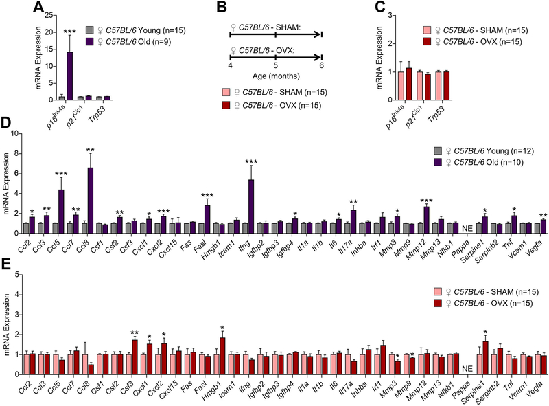
Fig 2. Comparisons of changes in senescence biomarkers and senescence-associated secretory phenotype (SASP) factors in osteocyte-enriched bone samples from female mice in the context of natural chronological aging and sex steroid deficiency.
(A) rt-qPCR analysis of in vivo age-associated changes in mRNA expression of the senescence effectors, p16Ink4a (Cdkn2a), p21Cip1 (Cdkn1a), and p53 (Trp53), in osteocyte-enriched bone samples from young (6-month-old, n=15) versus old (24-month-old, n=9) female C57BL/6 wild-type (WT) mice (data reproduced from J Bone Miner Res 31(11):1920–9, 2016). (B) Experimental design for testing the effects of ovariectomy-induced sex steroid deficiency on biomarkers of cellular senescence in bone; 4-month-old female C57BL/6 WT mice were randomized to undergoing either sham surgery (SHAM, n=15) or ovariectomy (OVX, n=15) for 2 months; all animals were sacrificed at age 6 months. (C) rt-qPCR analysis of in vivo changes in mRNA expression of the senescence effectors, p16Ink4a (Cdkn2a), p21Cip1 (Cdkn1a), and p53 (Trp53), in osteocyte-enriched bone samples from SHAM (n=15) versus OVX (n=15) female C57BL/6 WT mice. (D) rt-qPCR analysis of in vivo age-associated changes in mRNA expression of 36 established SASP factors in osteocyte-enriched bone samples from young (6-month-old, n=15) versus old (24-month-old, n=9) female C57BL/6 WT mice. (E) rt-qPCR analysis of in vivo changes in mRNA expression of 36 established SASP factors in osteocyte-enriched bone samples from SHAM (n=15) versus OVX (n=15) female C57BL/6 WT mice. Data represent mean ± SEM (error bars); NE = Not expressed (Cycle threshold [Ct] values >35). *p < 0.05; **p < 0.01; ***p < 0.001 (independent samples t-test).

