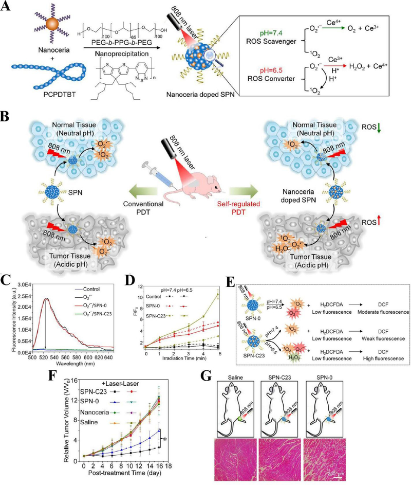
Figure 2.
Schematics show the components of SPNs doped with nanoceria and the concept of controllable photodynamic capacities of SPNs that are dependent on tissue microenvironments (A), and the differences between conventional PDT and self-regulated PDT when nanoceria are doped in SPNs (B). (C) In vitro studies show the capabilities of ROS scavenging by SPNs. Fluorescence intensities of ROS indicator were measured after addition of SPNs doped with different amount of nanoceria. (D) In vitro ROS generation from SPNs. The fluorescence changes of ROS indicator mixed with SPN-0 or SPN-C23 in different pH conditions irradiated with laser at 808 nm (0.44 W/cm2) as a function of irradiation time. (E) The graph illustrates the different responses of ROS in SPN-0 or SPN-C23 after irradiated with NIR laser and ROS responses were measured by H2DCFDA. (F) Tumor growth after mice were treated with different drug formulations. (G) Histological H&E staining of mouse muscles 24 h after the different treatments irradiated with NIR laser for 5 min. Reproduced with permission (Zhu et al., 2017). Copyright 2017, American Chemical Society.

