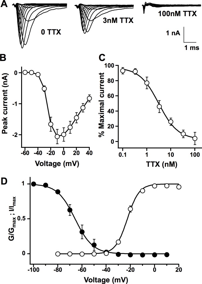
Fig 1. Endogenous sodium currents of ND7/23 cells.
(A) Families of sodium current traces recorded from untransfected ND7/23 cells under perfusion of external solution without (left) and with 3nM (middle) and 100nM (right) TTX. Calibration: 1nA, 1ms. (B) Current-voltage relation of the sodium current elicited by 100 ms square test pulse from –80 to +50mV in 10mV steps preceded by –120mV prepulse for 150ms and -70mV holding potential (means±standard error, n = 15). (C) TTX inhibition curve for the sodium current. Each data point was acquired using step depolarization to 0mV from –120mV and normalized to peak sodium current elicited under control perfusion prior to TTX application (means±SD, n = 3~11). IC50 was 3.3±1.0nM. (D) Conductance–voltage (open circle) and voltage-dependence of the steady-state inactivation (filled circle) relationships of ND7/23 cell sodium currents. Steady state inactivation used test pulse to 0mV, preceded by 500ms long prepulses to the indicated potentials and the holding potential was –120mV.

