
Fig 5. Total β-tubulin protein levels in Tubb2a and Tubb2b mutants.
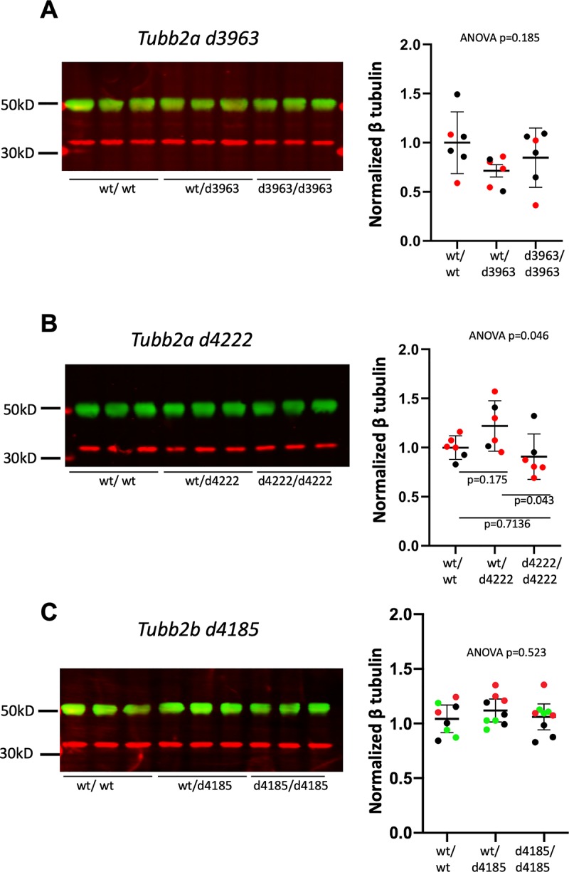
Western immunoblotting with an antibody recognizing multiple isoforms of β-tubulin for P0 brain lysates from (A) Tubb2ad3963 (B) Tubb2ad4222and (C) Tubb2bd4185 mice. Representative immunoblots for each allele are shown. Quantifications are of multiple experiments normalized to wild-type littermate controls. (A,B) Colors indicate littermates. n = 3 animals of each genotype, each set was run in duplicate. (C) Colors indicate replicate experiments from one set of littermates (Green bands indicate tubulin and red indicate GAPDH loading control for all immunoblotting experiments).