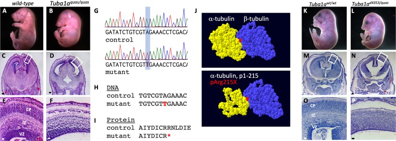
Fig 8. The quasimodo mutation is an allele of Tuba1a.
(A-F) Homozygous quasimodo mutants are distinguishable at E18.5 by the systemic edema (B). Histological analysis (E17.5) shows enlargement of the third ventricle (D) and reduced cortical tissue (F). (G) Sanger sequencing confirms homozygosity for a candidate SNP in a conserved DNA (H) and protein (I) sequence (SNP and coding change shown in red, red asterisk indicates premature stop codon). (J) Structure of the wild-type α/β tubulin dimer is shown above and the remaining α-tubulin structure is shown below (red = non-hydrolyzable GTP at the monomer interface). (K-P) Complementation analysis at E17.5 with the Tuba1ad4304 is consistent with quasimodo being an allele of Tuba1a as Tuba1ad4304/quas mutants have gross (L) and histological (N,P) phenotypes similar to the other Tuba1a homozygous phenotypes. The failure of alleles to complement indicates both mutations are in Tuba1a. Boxes in C,D,M,N show areas enlarged in E,F,O,P, respectively. Scale bars indicate 200 μm in C,D,M,N and 50 μm in E,F,O,P.

