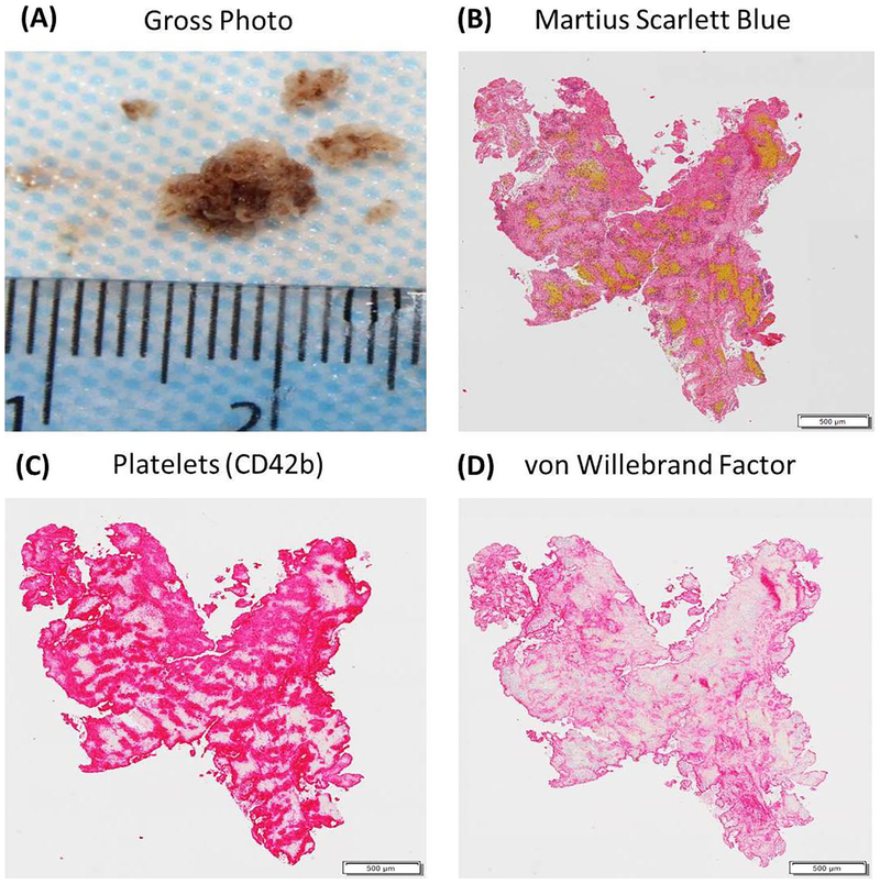
Figure 2: Histologic and Immunohistochemical Analysis of an acute ischemic stroke clot.
(A) is a gross photograph a ‘White’ clot retrieved from a patient using a mechanical thrombectomy procedure. (B) is an example of an MSB stained slide from the same clot showing the presence of Red Blood Cells (Yellow), White Blood Cells (Purple), Fibrin (Red) and Platelets/Other (Grey). (C) is an example of an immunohistochemically stained slide demonstrating the presence of Platelets in the clot (CD42b=Red). (D) is an example of an immunohistochemically stained slide demonstrating the presence of von Willebrand factor (Red).

