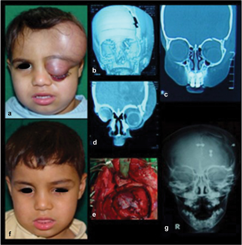
Fig. 7.

Extended isolated bone traumatic injury. ( a ) Frontal view shows expanding forehead, eyelids, and conjunctiva. ( b ) 3D-CT shows fracture of frontal and temporal bones and superior orbital rim. ( c, d ) Coronal CT shows fracture of orbital roof and superior orbital rim. ( e ) Coronal approach for exposure, dural repairs, and reduction and fixation. ( f ) One-year postoperative frontal view shows normal appearance. ( g ) Postoperative PA X-ray skull shows the reduced bones and fixation.
