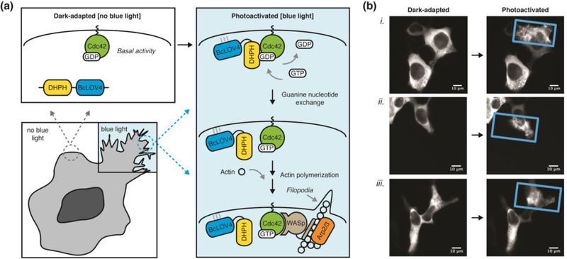
Figure 3. Spatially localized and optically induced cytoskeletal rearrangements by BcLOV4-derived opto-DHPH.
(a) Schematic overview of spatially precise opto-DHPH membrane recruitment and consequent activation of Cdc42 by the DHPH domain of the Intersectin GEF, which drives downstream actin polymerization. Cdc42 = Cell division control protein 42. DHPH = Diffuse B-cell lymphoma homology, Pleckstrin homology domain. GEF = Guanine exchange factor. Arp2/3 = Actin-related protein-2/3. WASp = Wiskott-Aldrich Syndrome protein. (b) Optically induced filopodia formation in HEK cells visualized by fluorescence imaging of a C-terminal mCherry tag. Only the blue light-illuminated (rectangle) regions show pronounced protusions, which are induced with very little stimulation (duty cycle = 0.8% = 0.5 sec per minute, λ = 450nm, 15 mW/cm2; spatially patterned by a digital micromirror device). Post-illumination times (i) 300 sec, (ii, iii) 500 sec Supplementary Movie 1 corresponds to cell (i).

