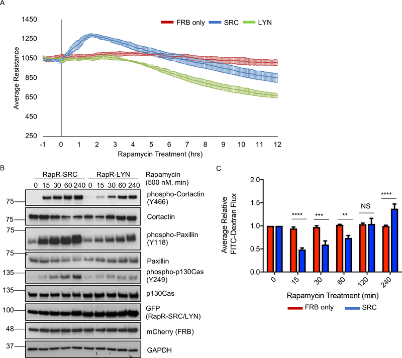
Figure 1: Effect of SRC activation on permeability of endothelial monolayer.
(A) TER analysis of HPAE cell monolayer. HPAE cells co-expressing cerulean-tagged RapR-SRC (SRC) and mCherry-FRB, cerulean-tagged RapR-LYN (LYN) and mCherry-FRB, or mCherry-FRB alone (FRB) were treated with rapamycin (500 nM) at time point 0. The graphs show the average resistance of 3 independent experiments and 90% confidence intervals. (B) Activation of RapR-SRC and RapR-LYN in HPAE cells. Cell lysates were collected at the designated time points and immunoblotted for the indicated proteins. (C) Fluorescein-labeled dextran flux across HPAE cell monolayer. HPAE cells co-expressing cerulean-tagged RapR-SRC and mCherry-FRB or mCherry-FRB only were treated with rapamycin (500 nM) to activate RapR-SRC. FITC-Dextran flux was measured at indicated time points before and after rapamycin treatment. The graph shows the averages and standard deviations from 4 independent experiments. Significance was evaluated using a two-way ANOVA with repeated measures and a post-hoc test with a Bonferroni’s multiple comparisons correction ***p < 0.001, **p < 0.01. All exogenous proteins were expressed using adenoviral transduction. See also figures S1A–E.

