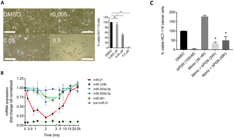
Figure 2: sub-cytotoxic concentration of 1 induces cellular proliferation arrest by modulating miR-21 expression in HCT-116 cells.
(A) HCT-116 cells were treated with DMSO, 0.005, 0.05, or 0.5 μM of 1 and incubated for 24 hr. Cells were then imaged, and viability was determined using the XTT cell-viability assay. Cell proliferation was inhibited at 0.05 μM and cell death was observed at 0.5 μM. ns = not significant, *p < 0.01, ** p < 0.001. (B) HCT-116 cells were treated with 50 nM 1 and total RNA was extracted from various time points (0–24 hr) for RT-qPCR analysis. Expression levels of miRs −21, −1246, −203a-3p, −200b-3p, and −361 and pre-miR-21 were analyzed using U6 snRNA as reference gene. miR-1246 did not show significant amplification up to 24 hr. (C) To show if miR-21 can rescue bPGN treated cells, HCT-116 cells were transfected with miR-21 mimic 4–5 hr before the cells were treated with 100 nM bPGN (2× GI50). Cell viability was measured at 24 and 48 hr by XTT assay. *p < 0.05. All experiments were performed in triplicate.

