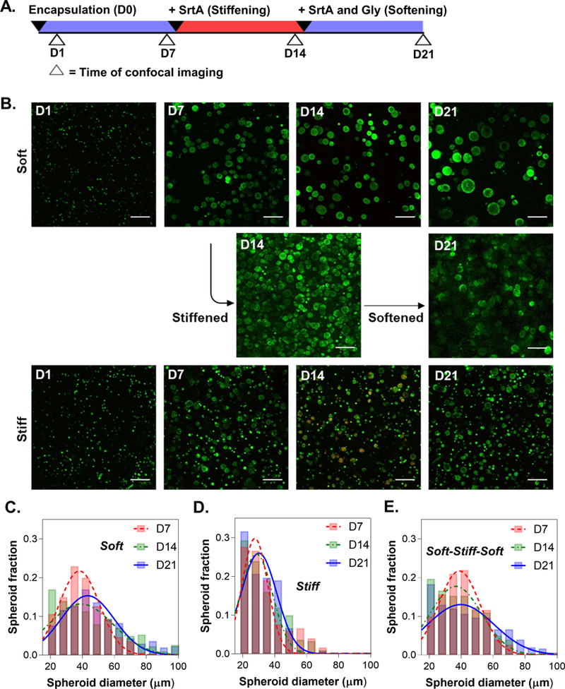
Figure 7.

(A) Timeline of alternate stiffening and softening of COLO-357-laden hydrogels. Hydrogels were stiffened on day 7 and softened on day 14. Time of confocal imaging is indicated by the open arrows. All imaging was completed prior to enzyme treatments (B) Representative confocal images of encapsulated COLO-357 cells in statically soft, stiff, and reversibly stiffened hydrogels. At least three z-stacked images per gel (10 slices, 100 μm thick) were taken. (Scale: 200 μm). Histogram of spheroids diameters for (C) non-dynamic soft, (D) non-dynamic stiff, and (E) reversibly stiffened and softened hydrogels (i.e., soft-stiff-soft).
