Figure 4.
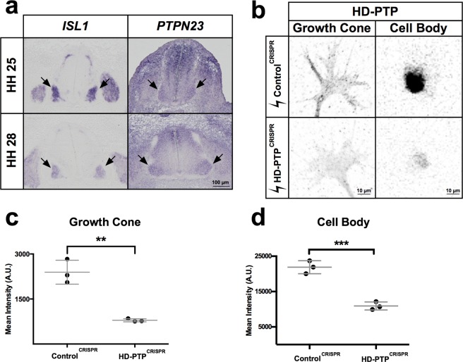
HD-PTP expression in embryonic motor neurons and CRISPR-mediated depletion. (a) Representative images of chick embryonic spinal cord sections at HH st. 25 and HH st. 28 where ISL1 and PTPN23 (chicken HD-PTP-encoding gene) mRNA was detected using in situ hybridisation. Note expression of PTPN23 in ISL1-expressing motor column (arrows). (b) Representative images of anti-HD-PTP antibody staining in growth cones and cell bodies of dissociated motor neurons harvested from embryonic spinal cords electroporated with ControlCRISPR or HD-PTPCRISPR plasmids. (c) Quantification of HD-PTP signals in growth cones of dissociated motor neurons harvested from embryonic spinal cords shows a decreased signal in HD-PTPCRISPR compared to ControlCRISPR (n = 3, 10–12 growth cones/n; p = 0.0023; Student’s t-test). (d) Quantification of HD-PTP signal in cell bodies of dissociated motor neurons harvested from embryonic spinal cords show decrease signal in HD-PTPCRISPR compared to ControlCRISPR (n = 3, 30–50 cell bodies/n; p = 0.0009; Student’s t-test). Values are plotted as mean ± SD. All values can be found in Supplementary Table S4. ***p < 0.001; **p < 0.01. Visible light images (a) and inverted grayscale fluorescent images (b).

