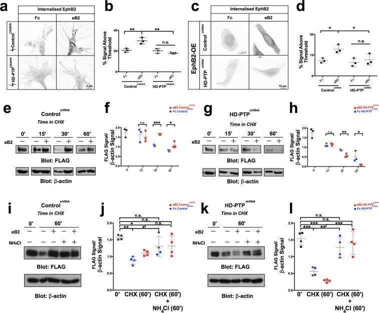
Figure 7.
HD-PTP loss increases the rate of EphB2 lysosomal degradation. (a) Representative images of ControlCRISPR and HD-PTPCRISPR spinal motor neuron growth cones, incubated for 20 min with 10 µg/mL eB2 or Fc and stained with anti-EphB2 antibodies followed by an acid stripping to show internalised EphB2. (b) Quantification of internalised EphB2 in ControlCRISPR and HD-PTPCRISPR motor neuron growth cones, incubated for 20 min with 10 µg/mL eB2 or Fc. ControlCRISPR growth cones showed a ligand-induced increase in internalised EphB2 (p = 0.0023), but HD-PTPCRISPR growth cones did not (p = 0.9831) (n = 3, 10–12 growth cones/n; one-way ANOVA followed by corrected Student’s t-tests). (c) Representative images of ControlshRNA and HD-PTPshRNA HeLa cells, incubated for 10 min with 1 µg/mL eB2 or Fc and stained with anti-EphB2 antibodies followed by an acid stripping to show internalised EphB2. (d) Quantification of internalised EphB2 staining in ControlshRNA and HD-PTPshRNA HeLa cells incubated for 10 min with 1 µg/mL eB2 or Fc. ControlshRNA showed an increase in internalised EphB2 signal upon eB2 stimulation (p = 0.0216), yet HD-PTPshRNA HeLa cells display no detectable increase in internalised EphB2 (p = 0.9001) (n = 3, 10–12 cells/n; one-way ANOVA followed by corrected Student’s t-tests). (e) Representative Western blot for EphB2 expression detected with anti-FLAG antibodies in transfected ControlshRNA HeLa cell lysates at different time points after incubation with 10 µg/mL protein synthesis blocker cycloheximide, exposed to either 1 µg/mL eB2 or Fc. β-actin detection is used as an internal control. (f) Quantification of Western blots for EphB2 detected with anti-FLAG antibodies in transfected ControlshRNA HeLa cell lysates after incubation with 10 µg/mL protein synthesis blocker cycloheximide together with either 1 µg/mL eB2 or Fc. FLAG signal intensity was normalised to β-actin and plotted for the different time points. By 30 min after cycloheximide treatment, eB2 stimulation appears to protect EphB2 from degradation compared to Fc (n = 3; Student’s t-test). (g) Representative Western blot for EphB2 detected with anti-FLAG antibodies in transfected HD-PTPshRNA HeLa cell lysates at different time points after incubation with 10 µg/mL protein synthesis blocker cycloheximide and either 1 µg/mL eB2 or Fc. β-actin is used as an internal control. (h) Quantification of Western blots for EphB2 detected with anti-FLAG antibodies in transfected HD-PTPshRNA HeLa cell lysates after incubation with 10 µg/mL protein synthesis blocker cycloheximide together with either 1 µg/mL eB2 or Fc. FLAG signal intensity was normalised to β-actin and plotted for the different time points. In contrast to ControlshRNA HeLa cells, in HD-PTPshRNA HeLa cells, eB2 stimulation appears to increase rate of EphB2 degradation compared to Fc by 30 min after cycloheximide treatment (n = 3; Student’s t-test). (i) Representative Western blot for EphB2 expression detected with anti-FLAG antibodies in transfected ControlshRNA HeLa cell lysates at different time points after incubation with 10 µg/mL protein synthesis blocker cycloheximide along with lysosomal blocker 10 mM NH4Cl, exposed to either 1 µg/mL eB2 or Fc. β-actin detection is used as an internal control. (j) Quantification of Western blots for EphB2 detected with anti-FLAG antibodies in transfected ControlshRNA HeLa cell lysates after incubation with 10 µg/mL protein synthesis blocker cycloheximide along with lysosomal blocker 10 mM NH4Cl, exposed to either 1 µg/mL eB2 or Fc. FLAG signal intensity was normalised to β-actin and plotted for the different time points. By 60 min after cycloheximide treatment and lysosomal blocking, EphB2 from degradation levels appear to be rescued (n = 4; Student’s t-test). (k) Representative Western blot for EphB2 expression detected with anti-FLAG antibodies in transfected HD-PTPshRNA HeLa cell lysates at different time points after incubation with 10 µg/mL protein synthesis blocker cycloheximide along with lysosomal blocker 10 mM NH4Cl, exposed to either 1 µg/mL eB2 or Fc. β-actin detection is used as an internal control. (l) Quantification of Western blots for EphB2 detected with anti-FLAG antibodies in transfected HD-PTPshRNA HeLa cell lysates after incubation with 10 µg/mL protein synthesis blocker cycloheximide along with lysosomal blocker 10 mM NH4Cl, exposed to either 1 µg/mL eB2 or Fc. FLAG signal intensity was normalised to β-actin and plotted for the different time points. By 60 min after cycloheximide treatment and lysosomal blocking, EphB2 from degradation levels appear to be rescued (n = 4; Student’s t-test). Values are plotted as mean ± SD. All values can be found in Supplementary Table S4. Full sized Western blots are in Supplementary Materials. CHX: cycloheximide; eB2: ephrin-B2-Fc; ***p < 0.001; **p < 0.01; *p < 0.05; n.s.: not significant. Inverted grayscale fluorescent images.

