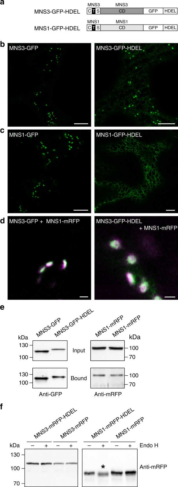
Fig. 5.

HDEL-mediated ER retrieval does not induce the redistribution of MNS3 to the ER. a Schematic illustration depicting the domain structure of the protein fusions MNS3-GFP-HDEL (full-length MNS3 C-terminally tagged with GFP and the ER retrieval signal HDEL) and MNS1-GFP-HDEL. b, c Confocal images (2 dpi) showing N. benthamiana leaf epidermal cells transiently expressing (b) MNS3-GFP or MNS3-GFP-HDEL, and (c) MNS1-GFP or MNS1-GFP-HDEL, respectively. Scale bars = 10 µm. d High-resolution Airyscan images of Golgi stacks from N. tabacum leaves labelled with MNS3-GFP (green) and MNS1-mRFP (magenta) or MNS3-GFP-HDEL (green) and MNS1-mRFP (magenta). Scale bar = 1 µm. e MNS3-GFP and MNS3-GFP-HDEL, respectively, were transiently co-expressed with MNS1-mRFP in N. benthamiana leaves. The GFP baits were purified using GFP-Trap beads and purified proteins were analyzed by SDS-PAGE and immunoblotting with anti-GFP and anti-mRFP antibodies. “Input” denotes total protein extracts before incubation with GFP-coupled beads; “bound” denotes the immunoprecipitated fraction. Source data are provided as a Source Data file. f Crude protein extracts from leaves of N. benthamiana ∆XF plants transiently expressing MNS3-mRFP-HDEL, MNS3-mRFP, MNS1-mRFP-HDEL, or MNS1-mRFP were subjected to Endo H digestion followed by immunoblotting with anti-mRFP antibodies. The asterisk marks the mobility shift. Source data are provided as a Source Data file
