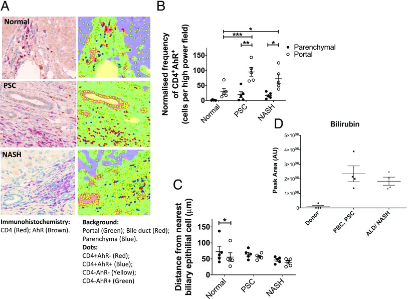
FIGURE 3.
AhR-expressing intrahepatic CD4 T cells are present at high density around bile ducts, and AhR ligand, bilirubin, is present in inflamed human liver tissue. (A) Localization of CD4+AhR+ and CD4+AhR− T cells in liver tissue examined by immunohistochemistry on paraffin-embedded liver tissue sections from normal, PSC, and nonalcoholic steatohepatitis (NASH) livers. Overlay of tissue segmentation and cell phenotype generated in inForm is displayed on the right. Original magnification ×20. (B) Frequency of CD4+AhR+ cells in parenchymal and portal regions of diseased and normal liver sections (n = 5) determined by immunohistochemistry analysis, normalized to the number of cells present in each tissue category per image. Significance was assessed by two-way ANOVA with Sidak (between locations [portal versus parenchyma]) and Tukey (between disease category) multiple comparisons post hoc tests. Significance intervals are indicated by asterisks (*). *p < 0.05, **p < 0.01, ***p < 0.001. Data are mean ± SEM. (C) Mean distance of CD4+AhR+ and CD4+AhR− cells from the nearest cell identified based on morphology was assessed by two-way ANOVA with Sidak (between cell types) and Tukey (between disease category) multiple comparisons post hoc tests. Significance intervals are indicated by asterisks (*). *p < 0.05. Data are mean ± SEM. (D) Bilirubin levels in normal liver and inflamed diseased livers were determined by UHPLC-MS. All data were normalized against the total ion count, and the statistical analysis was performed in R applying Mann–Whitney U test. ALD, alcoholic liver disease.

