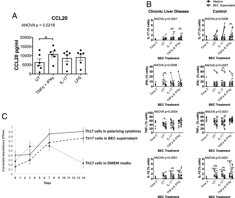
FIGURE 4.
LPS-exposed and inflamed BEC secrete CCL20, and culture of CD4 T cells in BEC-conditioned media promotes CD4 differentiation toward Th17 and maintains the survival of Th17 cells. (A) BEC were stimulated with soluble recombinant human cytokines (TNF-α + IFN-γ or IL-17) or LPS for 24 h, and the supernatants were analyzed for CCL20 chemokine secretion (n = 7). Effects of treatment were assessed by one-way ANOVA using Friedman nonparametric test with Dunn multiple comparisons post hoc tests (significant comparisons indicated by asterisks [*]) comparing the effects of each treatment to untreated control (UT). *p < 0.05. Data are mean ± SEM. (B) BEC were treated with type 17 cytokines for 24 h, and their supernatants were collected. CD4 T cells from chronic autoimmune liver disease patients and hemochromatosis (control) liver disease patients were stimulated with anti-CD3/CD28 beads and cultured in the BEC supernatants or control (recombinant cytokine containing media), and their expression of IL-17, TNF-α, IFN-γ, and IL-10 was examined at day 0 and day 7 by flow cytometry. Summary data for multiple donors are presented. Lines link frequencies of cytokine expression by cells from a given donor when cultured for 7 d in BEC supernatants versus the respective medium control. Effects of culture and treatment were assessed by one-way ANOVA using Friedman nonparametric test. Differences in expression compared with ex vivo were identified by Dunn multiple comparisons post hoc tests comparing to time 0 (significant comparisons indicated by crosses). Differential effects of BEC-secreted products compared with control medium at 7 d were identified by individual Wilcoxon matched pairs tests, applying the Bonferroni correction for multiple comparisons (significant comparisons indicated by asterisks [*] over a linking bracket). */+p < 0.05, **/++p < 0.01, ***/+++p < 0.001, ++++p < 0.0001. (C) Th17 cell viability over 14 d when cultured in supernatant from untreated BEC cultures (dashed line) was monitored and compared over 14 d to the viability of Th17 cells cultured in control medium (dotted line) or Th17-polarizing medium (IMDM supplemented with IL-1β, IL-6, and TGF-β) (solid black line); n = 3.

