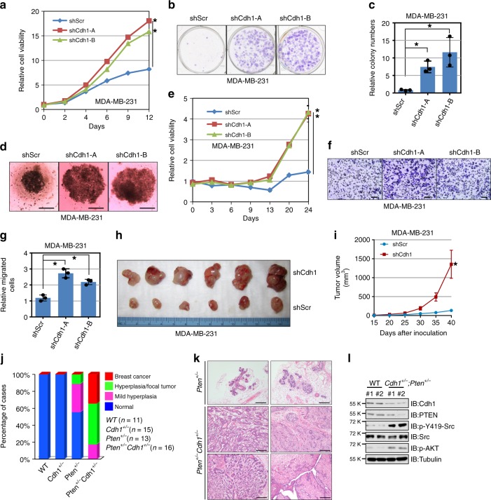
Fig. 2.
Cdh1 deficiency facilitates breast tumorigenesis. a MDA-MB-231 cells infected with control shRNA (shScr) or shCdh1 lentiviral constructs as described in Fig. 1b were subjected to cell proliferation assays in DMEM medium supplemented with 10% FBS for up to 12 days. Relative cell viability was determined at the indicated time points and was calculated as mean ± SD from three independent experiments. *P < 0.05; Student’s t test. b, c MDA-MB-231 cells generated in (a) were subjected to clonogenic survival assays in DMEM medium supplemented with 10% FBS for 14 days. Crystal violet was used to stain the formed colonies (b) and the colony numbers were calculated as mean ± SD (n = 3), *P < 0.05; Student’s t test (c). d, e MDA-MB-231 cells generated in (a) were subjected to 3D spheroid formation experiments in DMEM medium supplemented with 10% FBS for 24 days. The representative pictures are shown from three independent experiments (d), Scale bar, 100 μm. Anchorage-independent cell growth was measured using CellTiter-Glo 3D Cell Viability Assay Kit at the indicated time points (e). Relative cell viability was calculated as mean ± SD (n = 3). *P < 0.05; Student’s t test (e). f, g MDA-MB-231 cells generated in (a) were subjected to Transwell migration assays. The representative pictures are shown from three independent experiments. Scale bar, 100 μm (f). The relatively migrated cells were calculated as mean ± SD (n = 3). *P < 0.05; Student’s t test (g). h, i Tumor pictures (h) and the growth curves (i) for the xenograft experiments with the MDA-MB-231 cells generated in (a) were inoculated subcutaneously. In each flank of six nude mice, 3 × 10⁶ cells were injected. The visible tumors were measured at the indicated days. Error bars represent ±SEM (n = 6) (i). j The incidence of mammary gland hyperplasia and tumor in Pten+/− and Pten+/−;Cdh1+/− mice (6–7 months old) were quantified. k Hematoxylin and eosin (H&E)-stained sections of mammary tissues isolated from 6-month-old Pten+/− and Pten+/−;Cdh1+/− littermates. Scale bar, 100 μm. l Immunoblot (IB) analysis of lysates from breast tissues of Pten+/− and Pten+/−;Cdh1+/− mice

