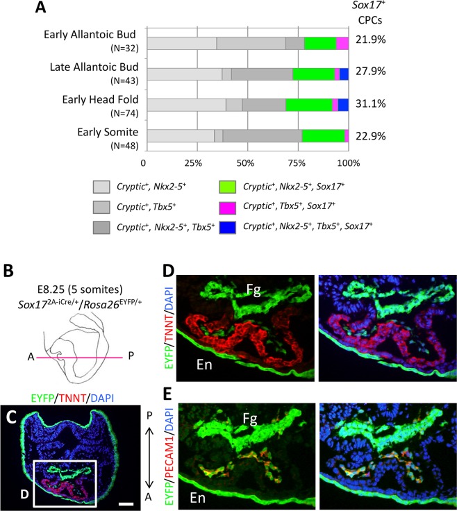
Figure 1.
Sox17+ CPCs specific for the endocardium differentiation. (A) Proportion of Sox17-expressing CPCs (Cryptic+, Nkx2-5+ and/or Tbx5+ cells) in mouse embryos at E7.5 (early allantoic bud, late allantoic bud, and early head fold stages) and E8.5 (early somite stage). N values indicate the number of cells examined. The data are derived from our previous study23. (B) Schematic representation of a mouse embryo at the five-somite stage (E8.25) as a left lateral view. The magenta line shows the sectional plane along the anterior (A)-posterior (P) axis in (C). (C–E) Immunofluorescence micrographs for EYFP (green), TNNT (red in C,D) and PECAM1 (red in E). Nuclei (blue) were stained with 4′,6-diamidino-2-phenylindole (DAPI). The boxed region in C is shown at higher magnification in D. The section shown in E is adjacent to that in D. Fg, foregut; En, endoderm. Scale bar, 100 µm.

