Figure 3.
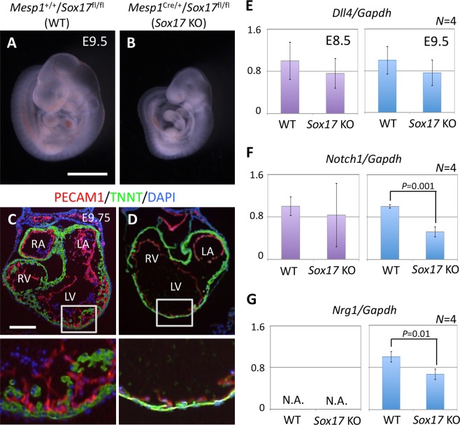
Cardiac defects associated with mesoderm-specific loss of function for Sox17 in mouse embryos. (A,B) Mesp1+/+/Sox17fl/fl (WT) (A) and Mesp1Cre/+/Sox17fl/fl (Sox17 KO) (B) embryos at E9.5. Scale bar, 1 mm. (C,D) Immunofluorescence micrographs in the heart of WT (C) and Sox17 KO (D) embryos at E9.75. The boxed regions in the upper panels are shown at higher magnification in the lower panels. Red, PECAM1; Green, TNNT; Blue, DAPI; LA, left atrium; RA, right atrium; LV, left ventricle; RV, right ventricle. Scale bar, 100 µm. (E–G) Reverse transcription and real-time PCR analysis of the relative expression levels for the NOTCH signaling-related genes Dll4 (E), Notch1 (F), and Nrg1 (G) in the heart of WT and Sox17 KO embryos at E8.5 (left panels) and E9.5 (right panels). Nrg1 expression at E8.5 was below the threshold for detection (N.A., not amplified). Only significant P values (Student’s t test) are indicated. Means ± SD.

