Fig. 1.
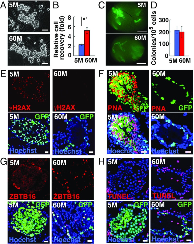
Long-term culture of GS cells. (A) Appearance of 5M-GS and 60M-GS cells. (B) Cell recovery after 5 d (n = 3). (C) Appearances of W recipient testes. (D) Colony counts (n = 8–12). (E–G) Immuno- and lectin staining of recipient testes with anti-γH2AX (E), PNA (F), or ZBTB16 (G) antibodies. (H) TUNEL staining of recipient testes 3 mo after transplantation. (Scale bars: A, 50 μm; C, 1 mm; E–H, 20 μm.) Asterisk indicates statistical significance.

