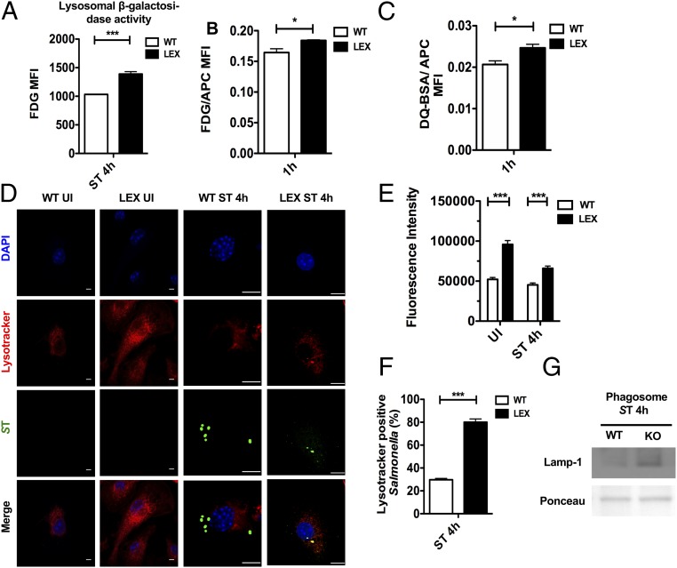
Fig. 5.
Reduced S. Typhimurium burden is mediated through enhanced phagolysosomal processing in Lepr-deficient macrophages. (A) Flow cytometric analysis of WT versus LEX BMDMs infected with C12FDG-coated S. Typhimurium 4 h after infection (n = 3). Flowcytometric analysis of WT and LEX BMDMs pulsed with C12FDG-coated beads (n = 3) (B) and DQ-BSA–coated beads (n = 3) (C). Bar graphs represent MFIs of C12FDG and DQ-BSA normalized to MFIs of red fluorescence. (D) Confocal microscopy analysis of UI and 4-h infected WT and LEX BMDMs pulsed with Lysotracker and stained for S. Typhimurium LPS. (E) Bar graph shows the fluorescent intensity of Lysotracker measured with ImageJ. (F) S. Typhimurium-LysoTracker colocalization in WT and LEX BMDMs 4 h after infection. (G) Lamp-1 expression on S. Typhimurium containing phagosomes 4 h after infection. Data are shown as mean ± SEM and statistical significance calculated using Student t test and represented as *P < 0.05; **P < 0.01; ***P < 0.001. (Scale bars: 20 μm.)

