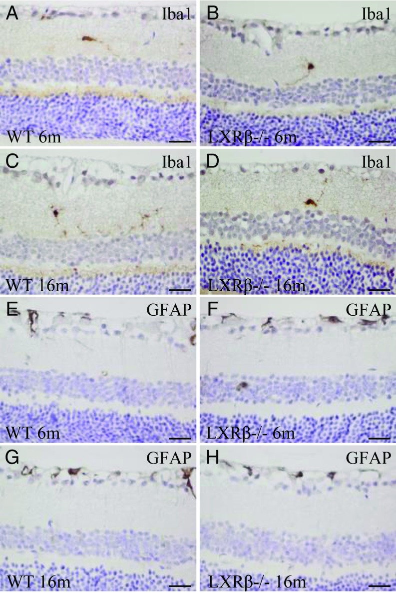
Fig. 3.
Microglia and astrocytes in the retina of LXRβ−/− mice. There were few microglial (Iba1-positive) cells in the retina of 6-mo-old WT or LXRβ−/− mice (A and B), and there was no increase in the number of these cells in16-mo-old mice of either genotype (C and D). At either 6 mo (E and F) or 16 mo (G and H) of age, there were few astrocytes in the retina, and there was no detectable difference between WT and LXRβ−/− mouse retinas. n = 4. (Scale bars: A–H, 20 μm.)

