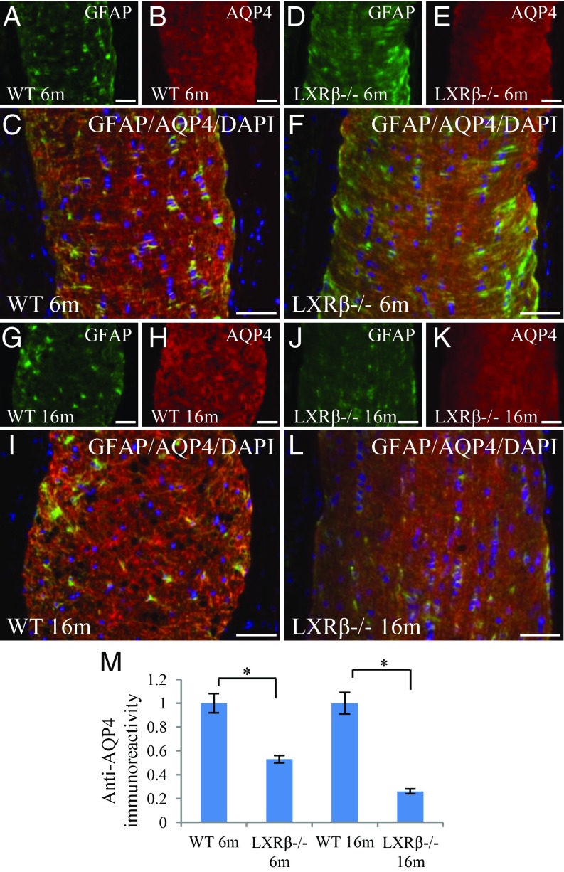
Fig. 5.
AQP4 in astrocytes in the optic nerve. In 6-mo-old WT mice, GFAP (green) and AQP4 (red) were strongly expressed at the feet and branches of astrocytes in the optic nerve (A–C). AQP4 expression was lower in the optic nerve of 6-mo-old LXRβ−/− mice (D–F). In 16-mo-old mice, GFAP and AQP4 were still strongly expressed in the optic nerve of WT mice (G–I), but their expression was reduced in the optic nerve of LXRβ−/− mice (*P < 0.05) (J–M). GFAP (green) was colocalized with AQP4 (red); the colocalized color is orange. The nuclei are counterstained with DAPI (C, F, I, and L). n = 4. (Scale bars: A–L, 50 μm.)

