
Figure 4.
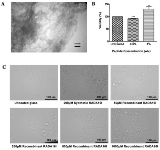
A) Transmission electron microscopy (TEM) image of RADA16I fibrils formed at a final peptide concentration of 2.5 mg.L-1. B) Evaluation of cell cytotoxicity of purified RADA16I fibrils. Blank, cells not treated with RADA16I; green, cells treated with 0.5% RADA16I; violet, cells treated with 1% RADA16I. C) Evaluation of cell adhesivity of purified RADA16I fibrils. i, uncoated glass cover slips; ii, glass cover slips coated with 200µM synthetic RADA16I; iii, glass cover slips coated with 50µM recombinant RADA16I; iv, glass cover slips coated with 250µM recombinant RADA16I; v, glass cover slips coated with 500µM recombinant RADA16I; vi, glass cover slips coated with 1000µM recombinant RADA16I.