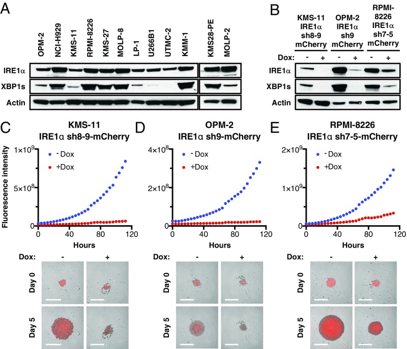
Fig. 1.
Expression of IRE1α in MM cell lines and effect of its depletion on spheroid 3D growth. (A) Twelve human MM cell lines were analyzed by immunoblot (IB) for protein levels of IRE1α and XBP1s. (B–F) KMS-11, OPM-2, and RPMI-8226 MM cells were stably transfected with a plasmid encoding doxycycline (Dox)-inducible shRNAs against IRE1α together with a plasmid encoding mCherry. Cells were incubated in the absence or presence of Dox (0.5 μg/mL) for 3 d, seeded on ultralow adhesion (ULA) plates, centrifuged to form single spheroids, and analyzed by IB for indicated proteins (B) or for growth based on mCherry fluorescence using an Incucyte instrument. (C–E, Lower) Representative images of indicated MM cells grown as single spheroids in ULA plates. (Scale bars, 800 μm.)

