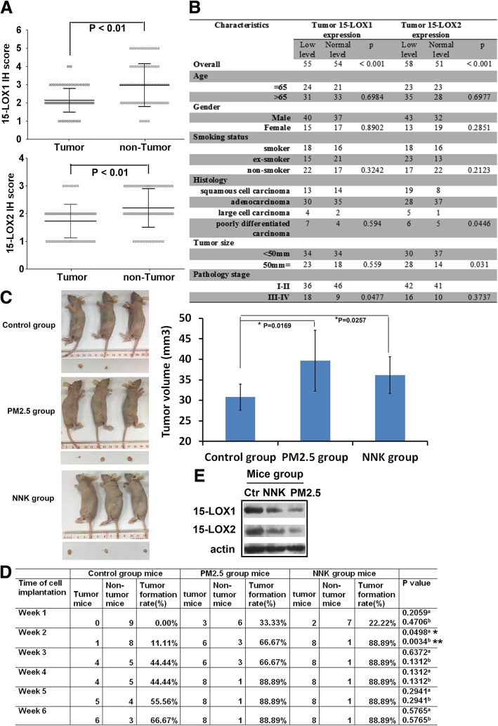
Fig. 3.
The decreased expression of 15-LOX1 and 15-LOX2 in lung tumor tissues. a The levels of 15-LOX1 and 15-LOX2 in 109 NSCLC tissues and paired adjacent normal tissues. The stained tissues were examined and expressed as Mean with range. Wilcoxon signed ranks test was used to compare the values (P < 0.01). b Baseline demographic characteristics of 109 human NSCLC patients underwent 15-LOX1 and 15-LOX2 analysis. The clinic-pathologic features in patients with relative expressing 15-LOX1/15-LOX2 were compared using Pearson’s chi-squared test or Fisher’s exact test for categorical variables. P < 0.05 was considered statistically significant. c Tumorigenicity assay. The nude mice were transplanted with NCI-H23 cells treated with PM2.5 or NNK for 28 days. Representative images of mice and tumor were shown. The growth of tumors was calculated by tumor volume (*P < 0.05). d Tumor formation rate assay. Tumor formation rate in each time-point was recorded (*p < 0.05, **p < 0.01; a P value as compared each group of PM2.5-treated condition with the control group; b P value as compared each group of NNK-treated condition with the control group. Data are mean ± SD). e 15-LOX1 and 15-LOX2 expression in the xenografts of NCI-H23 cells. Three of each group of tumor tissue proteins from week 6 mice was pooled together and 15-LOX1/15-LOX2 expression in the xenografts was detected. Equal loading was confirmed by probing with antibodies against actin

