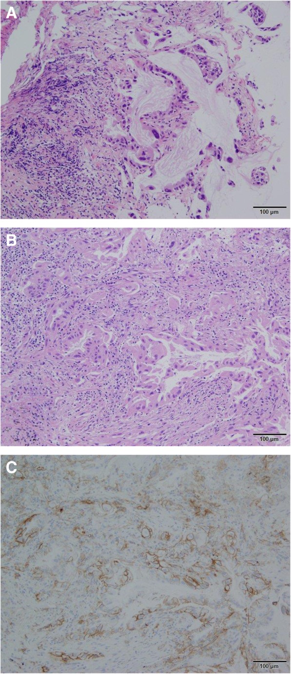
Fig. 2.

Representative image of HE staining for TBB and Cryo and PD-L1 ≥ 50% with the same patient (Adenocarcinoma 10×). a, HE staining for TBB specimens. b, HE staining for Cryo specimens. c, PD-L1 ≥ 50% for Cryo specimens. HE, hematoxylin and eosin; PD-L1, programmed death ligand 1; TBB, transbronchial biopsy; Cryo, Cryobiopsy
