Fig. 3.
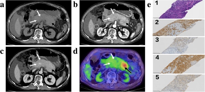
Type 1 autoimmune pancreatitis (AIP) with concurrent pancreatic solitary extramedullary plasmacytoma (SEP). Several areas of non-mass-like low-density (white and black arrows) in pancreas were found on arterial phase CT image (b). They appeared hypoattenuating on pre-enhanced (a) and venous (c) phase CT images. It was hard to differentiate SEP (black arrow) from the underlying AIP (white arrow) based on CT imaging findings (a, b and c). Fused fluorine 18 fluorodeoxyglucose (FDG) positron emission tomography/CT (18F-FDG PET/CT) (d) showed focal avid FDG uptake (black arrow) in the background of moderate FDG uptake (white arrow), which indicated there were two different entities present in the pancreas. The second biopsy of pancreas in the surveillance period showed dense infiltration of plasmacytoid cells on haematoxylin-eosin stain, original magnification × 200 (e-1). The plasmacytoid cells showed positive staining for CD138 (e-2), CD38 (e-3) and monotypic k-light chain (e-4), and negative staining for monotypic λ-light chain(e-5) on immunohistochemical stain, original magnification × 200

