
Figure 1.
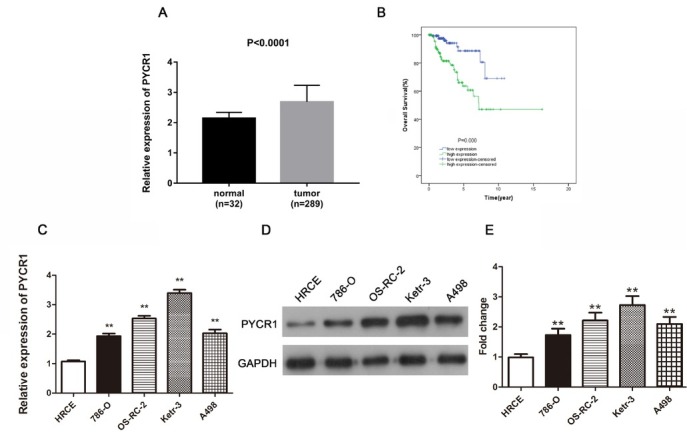
PYCR1 is promoted in PRCC tissues and cells, meanwhile up-regulated PYCR1 is related to poor prognosis in PRCC patients. (A) The relative expression of PYCR1 in PRCC tissues compared to normal tissues. (B) The overall survival curve in PRCC patients with high and low expression of PYCR1. (C) and (D) Comparison of PYCR1 expression in diverse cell lines through qRT-PCR and western blot analyses. (E) The quantification of protein expression level. All the data are expressed as mean ± SD and each test was performed in three times. (**P < 0.01). Supplement Figure 1: The correlation between PYCR2/PYCR3 expression and PYCR1 expression or survival rates of PRCC patients. (A) PYCR2 was over expressed in PRCC tissues consisting of 32 normal cases and 289 PRCC cases. (B) Survival curve showed the connection between PYCR2 expression and PRCC. (C) PYCR3 was increased in PRCC tissues including 32 normal cases and 289 PRCC cases. (D) The survival curves analysis on the basis of PYCR3 expression. (E) and (F) PYCR1 expression was closely linked with PYCR2/PYCR3 by the Pearson’s analyses.