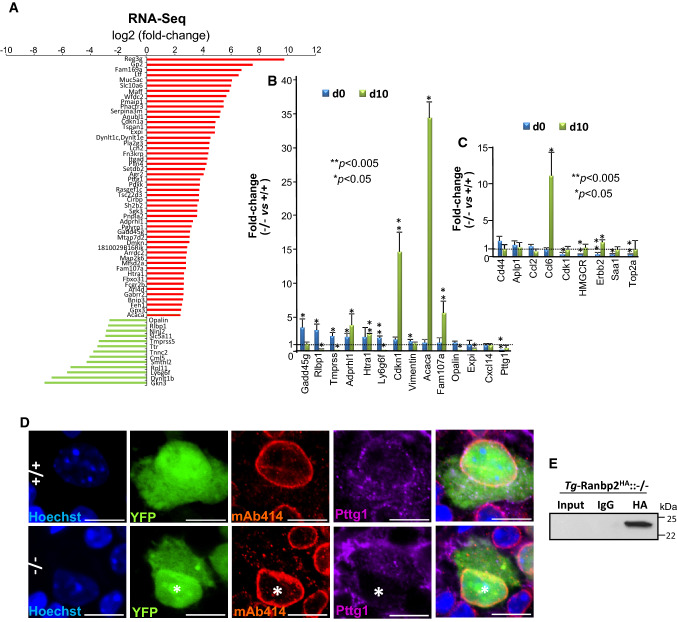
Fig. 5.
Differential transcriptome and gene expression analyses of optic nerve of retinal ganglion neurons (RGNs) upon loss of Ranbp2. A Differential RNA-Seq-based whole-transcriptome analysis of optic nerves between SLICK-H::Ranbp2flox/flox (−/−), SLICK-H::Ranbp2+/+ (+/+) and TgRBD2/3*-HA::SLICK-H::Ranbp2flox/flox at day 10 post-tamoxifen administration. Fifty transcripts were up-regulated (red) and thirteen transcripts were down-regulated in −/− mice at d10. B Validation of ranked RNA-Seq dataset by RT-qPCR and temporal and directional changes of expression levels of mRNAs between −/− and +/+ mice. Thirteen transcripts were validated to be up-regulated and Pttg1 was validated to be down-regulated by RT-qPCR in −/− mice at d0, d10 or both. Acaca (acetyl-CoA carboxylase alpha; also known as Acc1) and cdkn1 (cyclin-dependent kinase inhibitor 1) had the strongest up-regulation (~ 35 and 15-fold, respectively) at d10, whereas Pttg1 was down-regulated by ~ 25-fold at d0. Data are expressed as mean ± SD. Student’s t test, n = 3–4 mice/genotype. C Temporal and directional changes of expression of mRNAs in the optic nerve between -/- and +/+ mice and whose levels are known to be changed in the sciatic nerve by loss of Ranbp2. Five genes (Ccl6, Cdk1, HMGCR, erbb2, Saa1 and Top2a) were found also to be dysregulated in the optic nerve on d0, d10, or both. Ccl6 had the strongest change of expression (~ 11-fold increase). Data are expressed as mean ± SD. Student’s t test, n = 3–4 mice/genotype. D Confocal images of retinal flat mounts (ganglion neurons facing up) co-immunostained for Ranbp2 (Nup358)/Nup153/Nup62 (mAb414) and Pttg1. Pttg1 localizes at the nuclear rim with Ranbp2 (Nup358)/Nup153/Nup62 in YFP+-RGNs of +/+ mice, while Pttg1 localization is lost at the nuclear rim of YFP+-RGNs (nuclei labeled with *) of −/− mice. Scale bar 5 μm. E HA-tagged Ranbp2 (Tg-Ranbp2HA) expressed in transgenic mice with a null Ranbp2 background (−/−) co-immunoprecipitates Pttg1 from retinal extracts. Pttg1 is not detected in an overloaded aliquot of input extracts (first lane) owing to its very low abundance in retinal extracts. −/− SLICK-H::Ranbp2flox/flox, +/+ SLICK-H::Ranbp2+/+, Tg-Ranbp2HA:: −/− Tg-Ranbp2HA::Ranbp2−/−, d0 and d10 days 0 and 10 post-tamoxifen administration, respectively

