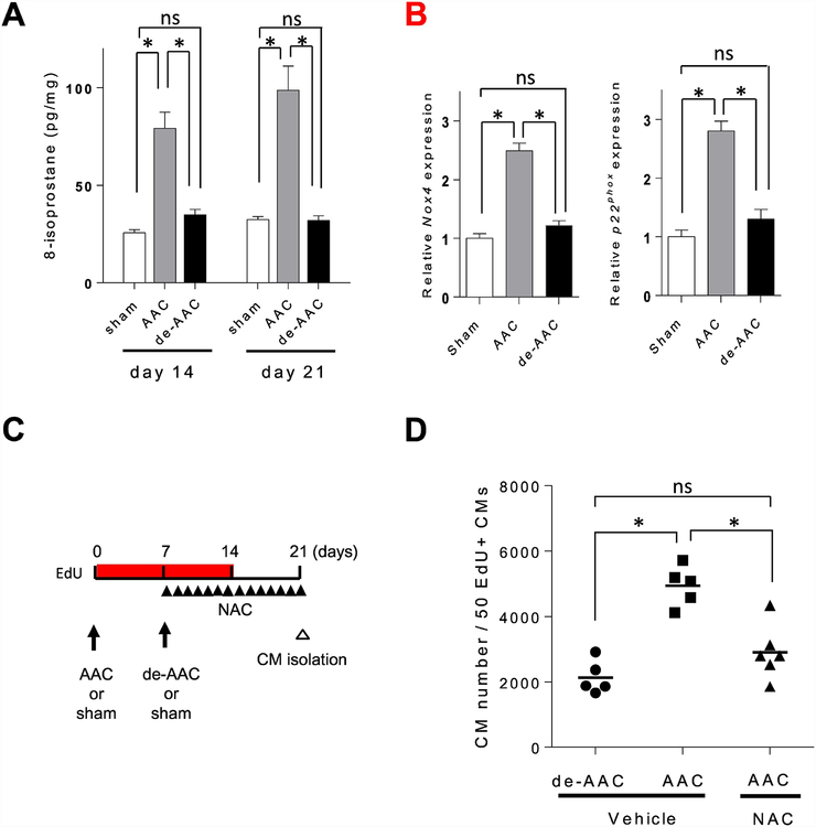
Figure 7. Abscising from oxidative stress after de-AAC contributes to cardiomyocyte proliferation.
A. The levels of oxidative stress in cardiac specimens were analyzed by 8-isoprostane assay. The levels of 8-isoprostane were increased in AAC heart in both postoperative day 14 and day 21, which were decreased to sham operation group levels after de-banding (sham; n = 3, AAC; n = 4, de-AAC; n = 4, *p<0.05). B. mRNA expression levels of NADPH oxidase subunits Nox4 and p22phox were increased in AAC compared to sham at postoperative day 814, and were comparable in de-banded heart (sham; n = 3, AAC; n = 4, de-AAC; n = 4). C. Study timeline. D. Total number of cardiomyocyte to find 50 EdU+ cardiomyocytes was increased in AAC heart compare to de-AAC heart, which was canceled by anti-oxidative agent, NAC, injection (de-AAC; n = 5, AAC/Vehicle; n = 5, AAC/NAC; n = 6, *p<0.05).

