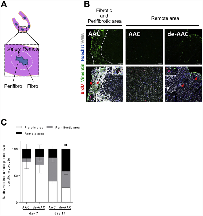
Figure 8. De-AAC induces further cardiomyocyte regeneration in LV area distant from injury.
A. Illustration of examined LV areas in AAC and de-AAC heart sections. B. Representative pictures from AAC (left and middle) and de-AAC (right) heart sections. Left panels show massive fibrotic and peri-fibrotic areas. C, Higher numbers of thymidine analog positive cardiomyocytes were observed in remote areas of de-AAC hearts compared to AAC. Error bars indicate SED for remote area, *p <0.05 vs AAC at 2nd week.

